Direct Current (DC) Cardioversion
This page is to inform you about your planned Direct Current (DC) electrical cardioversion. A cardioversion is an electrical treatment which aims to treat abnormal heart rhythms. It involves connecting you to a defibrillator machine and giving you a controlled electric shock. The procedure will involve you being admitted to Gloucestershire Royal Hospital as a day case. You will need to be collected from the hospital after the procedure and have someone at home with you overnight.
On this page
-
What does a cardioversion involve?
-
Potential risks of cardioversion
-
On the day of your procedure
-
After the procedure
-
Following your discharge
-
The normal heartbeat
-
What is Atrial Fibrillation (AF)?
-
What causes AF?
-
Risks of AF
-
Aims of the treatment
-
Why do I need an electrical cardioversion?
-
What are the benefits of cardioversion?
-
Are there any alternatives to cardioversion?
-
What will happen if I decide not to have the procedure?
-
Glossary of terms
-
Contact information
-
Further information
Before we can arrange for you to have a cardioversion, you will need to:
- Be taking an oral anticoagulant (OAC) (warfarin or one of the non-vitamin K oral anticoagulant medications such as Rivaroxaban, Dabigatran, Apixaban or Edoxaban.)
- If you are taking warfarin you will need to have a therapeutic International Normalised Ratio (INR) test (above 2.0) for at least 4 consecutive weeks before the date of your cardioversion.
- You should contact the Cardioversion Waiting List Coordinator on 0300 422 6543 as soon as possible if you are pregnant or have a cardiac pacemaker.
- If you have diabetes, you should contact the Cardioversion Waiting List Co-ordinator so that you can be advised about your medication or insulin for the day of the procedure.
What does a cardioversion involve?
As already discussed, a cardioversion is performed as a day case. Once you have been admitted, you will be asked to change into a hospital gown (this should be put on with the opening at the front, like a coat). An ECG will be performed.
You will be seen by a member of the Cardiology team, who will make sure that you are well enough to receive sedation for the procedure. The risks will be explained, and you will be asked to sign a consent form.
The cardioversion is performed in a quiet room. We will connect a blood pressure monitor, ECG electrodes and a finger probe to allow us to monitor your oxygen levels during the procedure.
A small plastic tube, known as a cannula, will be inserted into a vein on the back of your hand, or in your arm. The sedation will be given through the cannula. You will be asked to breathe some oxygen through a face mask. You will gradually drift off to sleep.
While you are asleep, a small electrical current will be passed through the chest using the defibrillator via 2 pads that will be stuck (temporarily) to your chest.
When the procedure has been performed, you will be taken back to your bed area to recover from the sedation.
Potential risks of cardioversion
DC Cardioversion is a safe and simple procedure, but we quote a risk of a complication in 1 in every 1500 procedures. Possible complications include:
- The procedure may not be successful – cardioversion is not 100 percent guaranteed to work. In many cases, patients go back to AF following a period of being in sinus rhythm, for others it is not possible to achieve sinus rhythm.
- Localised skin burning or chest wall discomfort – it is not uncommon for patients to experience some skin irritation following a cardioversion or some generalised aching in the chest itself. Sometimes, this will be just a little tenderness, other times there will be a pink mark on the skin where the pads have been on the skin. It is important to remember that this will resolve itself and that it is not a serious problem.
- Further rhythm disturbances – the aim of cardioversion is to correct the abnormal heart rhythm. However, sometimes when the electric current is passed through the heart, there is the potential to either cause a very slow heart rhythm which may lead to you needing a pacemaker or sometimes fast rhythms, originating in the ventricles, which require an additional shock to correct them. There is about a 1 in 1000 risk of needing a pacemaker to be fitted due to constantly slow heart rates following the cardioversion.
- Problems arising from a clot – this could be a stroke, a heart attack or a clot on the lung, also known as a Pulmonary Embolus (PE). In order to minimise this risk, we will have started you on an anticoagulant drug and (in the case of warfarin) you will be asked to have your INR monitored closely.
- If on the day of your cardioversion your INR is outside of your ‘target range’, we may have to postpone the procedure until a later date when it can be done safely.
- We will also postpone your procedure if you have missed any doses of your anticoagulant in the month before your procedure day.
On the day of your procedure
You must not eat or drink anything after midnight; this includes not chewing gum or sucking sweets.
The procedure will be performed by a member of the Cardiology team who will review you again before you are discharged.
After the procedure
- You will be taken to your bed area where you will receive care from a nurse trained to look after patients who have had sedation. A second ECG will be performed following your cardioversion.
- The nurses will record your pulse, blood pressure and oxygen saturations. They will also tell you when you can have a drink and something to eat and when it is safe to get out of bed.
- You will then be seen by a member of the Cardiology team who will discuss whether or not your cardioversion was successful. You will also be given any further information needed before being discharge.
Following your discharge
- You will need someone to pick you up from the ward and you must have someone at home with you overnight following your procedure.
- If this is not possible, please let the Cardioversion Waiting List Co-Ordinator know. The contact number is at the end of this page.
- You should not drive, operate heavy machinery, drink alcohol or sign important documents for 24 hours following your sedation.
- You should rest for a day or two before returning to your normal activities.
- Do not stop taking your anticoagulation medication and (in the case of warfarin) continue with INR monitoring, unless you are advised by a doctor. Only stop other cardiac medication if you are advised by your doctor or cardiac nurse. You may be at a higher risk of having complications if you stop taking medication without being advised to do so.
- You will be sent a follow up appointment to see your cardiologist after the procedure.
- If you go back into Atrial Fibrillation (AF) following discharge, and before your outpatient appointment, please contact your consultant secretary. Call hospital switchboard on 0300 422 2222
The normal heartbeat
The heart comprises of 4 chambers divided up according to the right and left sides of the heart. There are atria and ventricles. Blood entering the heart comes initially to the atrium on each side which serve as collecting chambers for the ventricles. The ventricles are the main pumping chambers of the heart and it is the blood expelled when the ventricles beat that you feel as a pulse.
The pumping action of the heart is controlled by electrical impulses which are produced in the right atrium in a cluster of special tissue known as the sinus node.
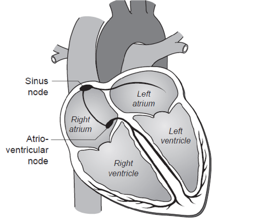
These activate the atria and cause the blood to be pushed down into the ventricles. The electrical impulses then pass down into the ventricles via another cluster of tissue called the Atrio-Ventricular node (AV node).
This works like a junction box and holds the signals up for a few moments before allowing conduction down the electrical fibres known as the Purkinje system. These activate the muscle in the ventricles which then contract and force blood out to the lungs and the rest of the body. The normal heart rate is dependent on many outside influences such as activity, state of health and medication. The normal heartbeat is called sinus rhythm.
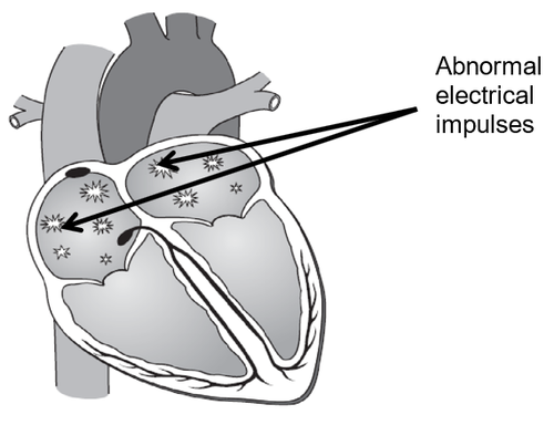
What is Atrial Fibrillation (AF)?
In AF, instead of the sinus node producing regular impulses, the atria beat very rapidly, effectively wobbling like a jelly.
Only a small number of these impulses can be conducted down through the AV node to the ventricles but this still produces ventricular rates much faster than normal.
Fast heart rates often lead to a variety of symptoms such as breathlessness, reduced exercise tolerance, chest pain, dizziness and lethargy
What causes AF?
A variety of triggers can cause AF. In some patients we are unable to find a cause but in others the cause could be disease of the heart valves, narrowing of the coronary arteries, poor function of the heart (for a variety of reasons), infections (especially lung infections or pneumonia), chronic lung disease or excess alcohol consumption.
AF is increasingly common as you get older and can happen in up to 1 in every 3 people over the age of 60. It can, however, happen at any age. Around 800,000 people in the UK have AF – roughly 1 in every 100 and mostly aged 55 or over.
AF is divided into 3 main categories:
- Paroxysmal – this happens intermittently with episodes lasting anything from a few seconds to many hours.
- Persistent – this is atrial fibrillation which has been recently discovered and attempts are being made to return the heart to its normal rhythm.
- Permanent – in which the heart cannot be returned to its normal rhythm and AF has been accepted.
Risks of AF
AF is often not immediately dangerous or life threatening but there are 2 main problems associated with it:
- Risk of stroke – As the rhythm of the heart is irregular; blood does not flow through the heart smoothly. As a result of this, clots of blood may form within the heart. These may flow out of the heart and cause a stroke. The risk of stroke is dependent on various factors such as age, high blood pressure and diabetes.
- Risk of heart damage – The heart has a tendency to run much faster than normal in AF. If this carries on for a prolonged period of time, the muscle wall of the heart can become damaged. This can itself lead to breathlessness and a condition known as heart failure.
Aims of the treatment
The main aims of the treatment are:
- To reduce the risk of having a stroke by the use of blood thinning medication known as anticoagulants.
- To reduce your heart rate by the use of rate controlling medication.
- To reduce your symptoms and improve your quality of life.
Why do I need an electrical cardioversion?
When you are in AF, your heart is unable to pump as efficiently. This can lead to palpitations and feeling breathless and tired. Some people may also experience chest pains, dizziness or feeling faint. In some patients, however, there are no symptoms and the irregular pulse or abnormal ECG is only noticed during a routine checkup.
AF increases the risk of blood clots developing in your heart. This is serious as it increases the risk of you having a stroke (loss of brain function as a consequence of interruption of the blood supply to the brain).
If your AF is caused by another medical condition, your doctor will usually recommend that this is treated first and they then may recommend a cardioversion.
What are the benefits of cardioversion?
The cardioversion is performed to improve your symptoms although it may also be possible for your doctor to discontinue some of your medications. Sometimes we perform cardioversion when the function of your heart has been reduced and we want to find out whether this is due to the AF and see if it improves by restoring normal heart rhythm.
Are there any alternatives to cardioversion?
Some symptoms of AF can be treated with heart slowing medication such as beta blockers. Anticoagulant medication can reduce the risk of having a stroke. However, medication may not be as effective as an electrical cardioversion in relieving your symptoms.
In some cases, catheter ablation treatment is given. This uses heat or cold to create scars inside your heart which can interrupt the abnormal electrical signals. Catheter ablation carries a higher risk than electrical cardioversion and may not be effective if you have had AF for a long time.
What will happen if I decide not to have the procedure?
You will be referred back to your cardiologist who may be able to recommend alternative treatment.
Glossary of terms
The information below will explain some of the abbreviations used on this page:
|
International Normalised Ratio (INR) |
The blood test used to calculate your warfarin dose |
|---|---|
|
Electrocardiograph (ECG) |
A trace showing the heart rhythm and electrical activity within the heart |
|
Atrial Fibrillation (AF) |
An irregular arrhythmia affecting the top chambers of the heart, the atria |
| OAC |
Oral anticoagulant – a blood thinner used to reduce the risk of stroke in AF |
Contact information
If you have any questions about your planned procedure, please contact the Cardioversion Waiting List CoOrdinator. However, if you have concerns about your existing treatment or any of the information on this page please contact your Cardiology Consultant’s secretary.
Cardioversion Waiting List Co-Ordinator
Tel: 0300 422 6543
Monday to Friday, 9:00am to 4:00pm
Hospital switchboard for cardiology consultant secretaries
Tel: 0300 422 2222
Further information
Arrhythmia Alliance
Helpline: 01789 867 501
Website: www.heartrhythmalliance.org
British Heart Foundation
Heart Helpline: 0808 802 1234
Website: www.bhf.org.uk
Atrial Fibrillation Association
Website: www.afa.org.uk
Helpline: 01789 867502